
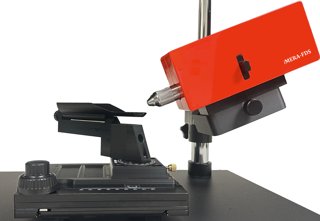
Fundus camera
High-resolution iMera-fundus camera engineered for preclinical retinal imaging in rodent models. Multi-modal, customizable, and built for longitudinal studies.
Field of view
Modalities
Animal model
Interface
Configure your system
Every lab is different. Let's build the right system for yours.
⏱ Typical lead time: 6–10 weeks after order
Multi-modal imaging
Color fundus and FA — with optional GFP, NIR, and ICGA
Image all species
iMera system covers mouse, rat, rabbit, cornea, dog, Zebrafish, and primate.
Longitudinal study ready
High dynamic 6-DOF stage enables reproducible positioning for tracking retinal changes
High-resolution imaging
1.4 µm/pixel resolution with 900×900 pixel output across up to 50° field of view.
Specification
Value
Fieldof view
Upto 50°
Image resolution
1.4μm/pixel (mouse)
Optical resolution
4.5μm (mouse)
Pixel resolution
900× 900 pixels
Imaging modalities
Color, FA
Optional modalities
GFP, NIR, ICGA (mono sensor & light source upgrade required)
Light source
High-power white & blue LED
Camera sensor
Scientific CMOS
Objective lens
One lens – mouse to primate
Animal stage
6-DOF motorized stage
Image format
TIFF, JPEG, PNG, AVI
Compatible species
Mouse, Rat, Rabbit, Dog, Primate
Acquisition software
iMera Capture
Warranty
1 year standard, extended available
Wavelengths, optics, software, hardware — built for your experiment.